A decade of collaboration between Bionirs Arg SA and physicists from the Universidad Nacional del Centro de Buenos Aires (UNCPBA) related to the propagation of light within biological tissues, specifically breast tissue, led to the development of an approach innovative and design of a clinical imaging device for the detection of breast cancer.
 How does MamoRef work?
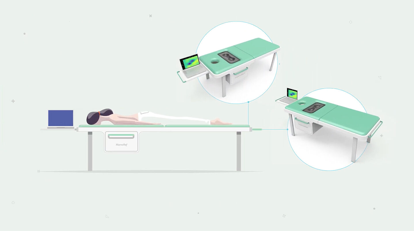
VERSIÓN 2.0

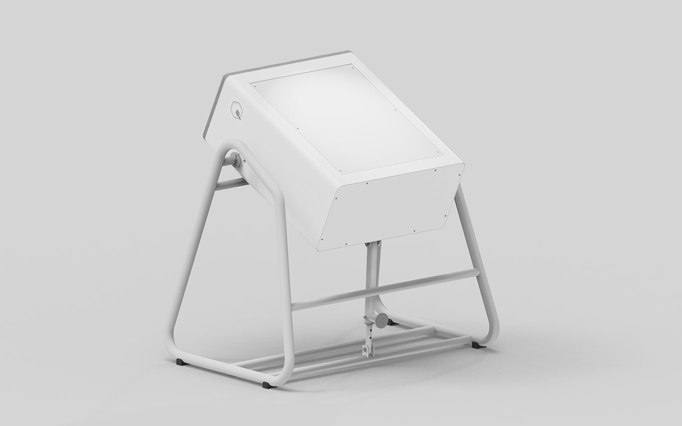
VERSIÓN 1.1

MAmoRef consists of a station with a transparent window under which the measurement system is placed. The patient sits on a chair and lies with her breasts resting on the window, avoiding any compression, allowing any woman to use it without limitation.
.

About NIR light
This NIR light is between 600 and 900 nm in wavelength, in the so-called “optical or therapeutic window” (Figure 2), and is poorly absorbed by the most significant components of soft tissue, thus being able to penetrate them by several centimeters and explore them. When light enters the tissue, it does not travel a straight path but diffusely scatters or “scatters”, part of it leaving the same face that entered the tissue (that is, it is “diffusely” reflected). The way in which this propagation occurs as well as the amount of light that is absorbed at each wavelength depends on the physiological and metabolic characteristics of the tissue traversed.
NIR light is known to provide valuable data on cell function and biological processes such as blood flow, hemoglobin concentration, and oxygen saturation. NIR imaging has been used to determine the concentration of oxygen saturation of hemoglobin in healthy and cancerous tissue.

Figure 2: Taken from Vogel2003

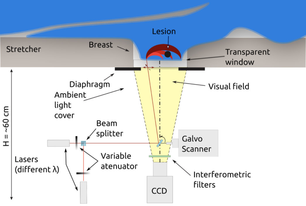
Schematic representation of MamoRef System.
Each pixel of each image is treated as an independent detector, thus there are thousands of source-detector pairs for each wavelength. Using a model of the most probable paths of light within the tissue, it is possible to infer which portion of the tissue affects each of these pairs and, for example, to be able to determine which areas of the breast have greater or lesser absorption of light in each one. wavelengths. Thus then, the images are processed with an algorithm that cleanses, and reconstructs a final image for clinical evaluation.
Based on the functional premise that the propagation of different wavelengths of light in the NIR region depends on the type of tissue, it is possible to obtain a map of oxygenation and hemoglobin concentration, related to metabolic function.
MamoRef has the potential to help in the diagnosis of complementary evaluation and classification of injuries in a simple, efficient and non-invasive way, providing metabolic information associated with the type of injury.
These uses are based on the following hypotheses to be clinically validated:
.
Scientific Work
- Simultaneous retrieval of optical and geometrical parameters of multilayered turbid media via state-estimation algorithms.
- GPU accelerated Monte Carlo simulation of light propagation in inhomogeneous fluorescent turbid media. Application to whole field CW imaging.
- Wide field continuous wave reflectance optical topography including a clear layer on top of the diffusive surface.
- Retrieval of the optical properties of a semi-infinite compartment in a layered scattering medium by single-distance, time-resolved diffuse reflectance measurements.
- An Improved Extended Kalman Filter algorithm for Diffuse Optical Tomography.
- A novelty technique for the fabrication of biomedical optics phantoms with cyst – mimicking inclusions.
- Photoacoustic Determination of Speed of Sound in Binary Mixtures of Water and Ethyl and Methyl Alcohol.
- Study of inks used in biomedical optics phantoms: stability and ageing
- CW Fluorescence imaging of tissue-like media in reflectance geometry
- Determination of the optical properties of multilayered phantoms by time-resolved reflectance measurements.
- Diffuse light transmission profiles obtained using CW: A comparative analysis with time resolved experiments.
- Diffuse reflectance optical topography: location of inclusions in 3D and detectability limits.
